UCT Clean Screen EtG Sorbent Cited in Nerve Agent Article
The use of chemical weapon nerve agents is banned by international convention and widely considered a human rights violation. Nonetheless, nerve agents have been used in attacks on military personnel and civilians in Iraq, Japan, Malaysia, Britain and Syria. For forensic and judicial purposes, verification of nerve agent exposure from biomatrices is needed to unambiguously identify the use of specific nerve agents following attacks. These agents belong to one of two groups: the G-agents or the V-agents. G-agents include Tabun (GA), Sarin (GB), Soman (GD) and Cyclosarin (GF).
A recently proposed model for the incorporation of xenobiotics of forensic interest into the human skeleton suggests nerve agent metabolites may incorporate into bone at relatively elevated concentrations based on their unique chemical properties. In a recent paper authored by Katie M. Rubin et al., UCT’s famous EtG solid phase (SPE) extraction cartridge was employed to test the hypothesis that nerve agent metabolites interact with bone. In this study methods for the extraction, isolation and semi-quantitative detection of nerve agent metabolites (MPA, EMPA, IMPA, iBuMPA, CMPA and PMPA, corresponding to the nerve agents VX, Russian VX, sarin, cyclosarin and soman, respectively) from osseous tissue were developed using LC-MS/MS and LC-TOF instrumentation.
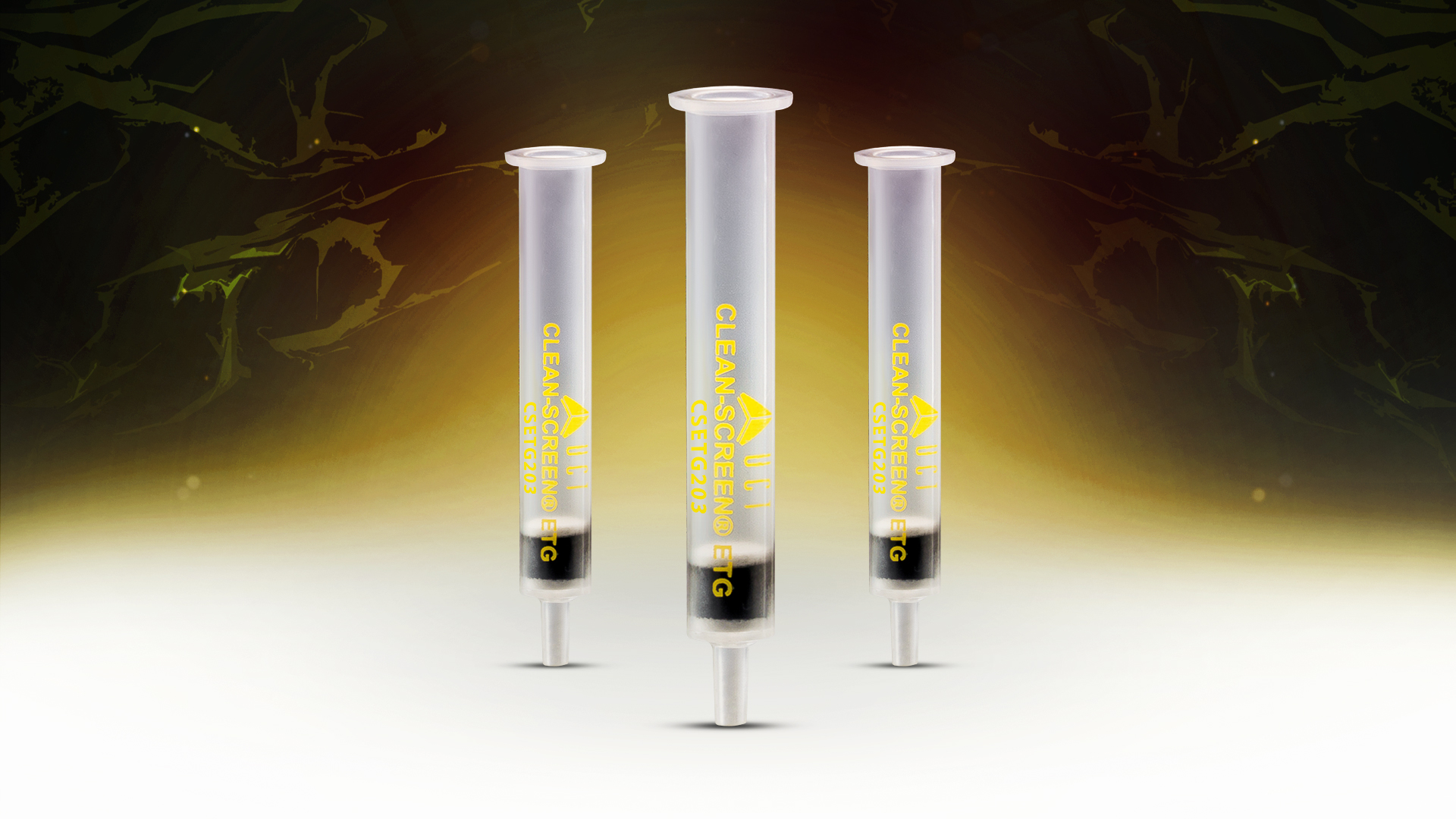
In the validated methodology, ETG cartridges (10 mL capacity, 400 mg proprietary, carbon-based sorbent) were conditioned with 4.0 mL 1% FA in MeOH, followed by 4.0 mL 1.2 M HCl. Samples underwent loading onto the SPE sorbent by gravity. Columns were rinsed with 2 × 4.0 mL 1.2 M HCl and then dried under full pressure for 10 min. Analytes were eluted with 1.0 mL 1% FA in MeOH. Eluates were dried under a gentle stream of nitrogen at 40◦C. Residues were reconstituted in 0.2 mL 0.5% AA in water and transferred to LC insert vials
The optimized methods were validated on the LC-MS/MS instrument. The achieved limits of detection (5–20 pg per g for four analytes; 350 pg per g for the fifth analyte) were lower than many of those published for the same analytes in other biomatrices, including serum and urine. These methods were tested on the skeletal remains of minipigs exposed to the chemical weapon VX in vivo. The VX metabolite was detected in multiple minipig bone samples.
The authors report that this is the first-time in vivo nerve agent exposure has been detected from bone. Further, detected concentrations and diaphyseal-to-epiphyseal area count ratios reflect animal exposure history. Although the results are limited, they are promising, indicating that nerve agent metabolites may interact with bone as a pharmacokinetic compartment and can be extracted from bone postmortem. Additional studies assessing the effects of different agents, exposure pathways and taphonomic variables, are needed; however, these results suggest the method may be used with human bone to detect use of chemical weapons from postmortem biomatrices even well after a suspected attack.
Click here for more information regarding our Clean Screen EtG columns and available configurations.